

Earlier particle insight, earlier understanding.
Subvisible particles tell us a story
We Give You the Data to Control
Visible and subvisible particles in parenteral products can trigger stability concerns, investigations, batch holds, and difficult QA decisions. Routine particle counts may show that particles are present — but they often do not explain what they are, where they came from, or whether they indicate a material, process, container-closure, or handling issue.
SizeID.bio provides particle-level evidence for development, stability studies, and investigations. We analyze large particle populations using deep-learning image classification and Raman-based chemical identification to characterize particle size, morphology, chemistry, and likely source.
This helps QA, analytical development, and manufacturing teams move from particle findings to source attribution, mechanism understanding, and CAPA-relevant conclusions.
Particle investigations begin with understanding what the particles are.
-
Identify foreign matter and recurring patterns early—before they enter production. Support supplier qualification, incoming inspection, and risk-based controls.
-
Monitor microbes and particulates together. Reveal biofilm-related shifts, corrosion signatures, polymer residues, and other contributors that simple counts can miss.
-
Explain excursions across cleanroom zones by differentiating typical contributors (skin, fibers, equipment wear, packaging residues) and highlighting emerging patterns.
-
Detect drift, equipment wear, cleaning issues, and process-related shifts through particle population changes and material attribution.
-
Quantify contributions from silicone, rubber, polymers, glass, and steel—supporting component risk assessments and investigation narratives.
-
Contextualize reject trends and long-term changes by tying "what is seen" to "what it is," enabling targeted remediation and comparability arguments.
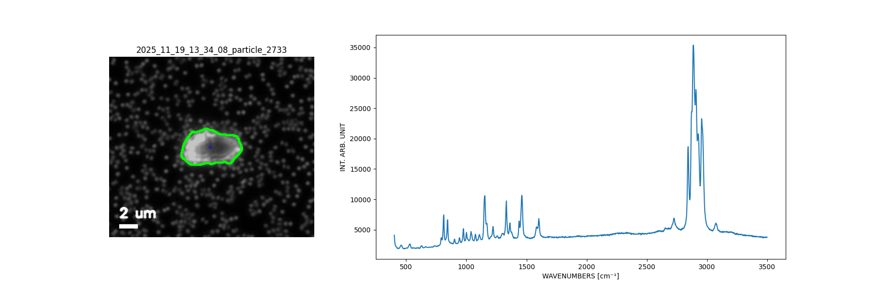
Tyvek
Tyvek (Stopper Bag)

Cellulose

Polystyrene